
Table of Contents
ToggleA Comparative Guide to Nursing Interventions, Monitoring, and Safe Administration
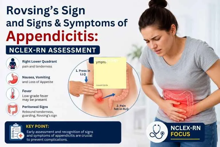
A patient has abdominal pain that began near the belly button and moved to the right lower quadrant. Pain on the right side when the left lower quadrant is palpated may indicate Rovsing’s sign, a finding associated with suspected appendicitis.
For NCLEX-RN students, it is important to know how Rovsing’s sign is assessed, what a positive finding suggests, and how it fits into the patient’s overall clinical picture.
Appendicitis may cause abdominal pain, nausea, vomiting, loss of appetite, and fever, although not every patient has all these symptoms. Recognizing these patterns can help with NCLEX-RN clinical judgment and prioritization questions.
What Is Rovsing’s Sign?
Rovsing’s sign is a physical examination finding associated with peritoneal irritation, especially when appendicitis is suspected.
A positive Rovsing’s sign occurs when palpation of the left lower quadrant (LLQ) causes pain in the right lower quadrant (RLQ). This may result from irritation of an inflamed appendix or surrounding peritoneal tissues.
However, Rovsing’s sign does not confirm appendicitis on its own. It must be considered along with the patient’s history, symptoms, laboratory findings, and, when needed, imaging.
What Does a Positive Rovsing’s Sign Indicate?
A positive finding suggests that appendiceal or peritoneal irritation may be present, especially when it occurs with other findings such as:
- Right lower quadrant tenderness
- Pain that migrated from the periumbilical area
- Nausea or vomiting
- Loss of appetite
- Fever
- Guarding or other signs of peritoneal irritation
For an NCLEX-RN question, think of Rovsing’s sign as supporting evidence, not a standalone diagnosis.
How Is Rovsing’s Sign Assessed?
During an abdominal examination, the examiner applies pressure or palpates the left lower quadrant and observes the patient’s response.
A positive finding is pain felt in the right lower quadrant during this maneuver.
The important NCLEX-RN point is not simply remembering “left side equals right-side pain.” You should understand what the finding means in context.
Remember: A positive Rovsing’s sign can increase suspicion for appendicitis, but a negative sign does not rule it out.
Signs and Symptoms of Appendicitis
Appendicitis occurs when the appendix becomes inflamed, usually as a result of an obstruction. If untreated, it can progress to rupture and peritonitis. Symptoms vary between patients, so the classic presentation should guide assessment but not be treated as a definitive diagnosis.
Abdominal Pain
Abdominal pain is the key symptom. A classic pattern is:
- Pain begins around the umbilicus.
- It becomes more severe.
- It travels into the right lower quadrant (RLQ).
- Pain becomes more localized.
- Movement, coughing, or jarring may worsen the pain.
RLQ tenderness is common, but pain location can vary based on appendix position, age, and other factors.
Nausea, Vomiting, and Loss of Appetite
Patients may experience nausea, vomiting, and anorexia (loss of appetite). These symptoms are not specific to appendicitis, so consider them alongside the pain pattern. For NCLEX-RN questions, remember that abdominal pain often occurs before vomiting in appendicitis.
Fever and Other Systemic Findings
A patient may develop low-grade fever, malaise, abdominal swelling, constipation, diarrhea, or difficulty passing gas. Laboratory tests may show leukocytosis (increased WBC count), but a normal WBC count does not rule out appendicitis, and an elevated count does not confirm it.
Physical Assessment Findings in Appendicitis
Physical Assessment Findings in Appendicitis
Rovsing’s sign is one of several physical findings that may support suspicion of appendicitis. These findings should always be interpreted along with the patient’s overall clinical presentation.
McBurney’s Point Tenderness
McBurney’s point is a classic area of tenderness in the right lower abdomen near the expected location of the appendix. Tenderness here may support suspected appendicitis, but anatomical variation means it is not present in every patient.
Rebound Tenderness
Rebound tenderness occurs when pain increases after pressure is released from the abdomen. It may indicate peritoneal irritation, but does not confirm appendicitis.
Abdominal Guarding
Guarding occurs when the abdominal muscles tighten during palpation. Voluntary guarding may result from anxiety or anticipated pain, while involuntary guarding can suggest peritoneal irritation.
Psoas Sign
The psoas sign is positive when movement that stretches or activates the psoas muscle causes RLQ pain. It may occur when an inflamed appendix is positioned near the psoas muscle, such as with a retrocecal appendix.
Obturator Sign
The obturator sign involves RLQ or pelvic pain during movement that stretches the obturator internus muscle. It may be associated with a pelvic appendix and should be considered alongside other clinical findings.
Rovsing’s Sign vs Other Appendicitis Signs
Finding | Assessment / Meaning |
Rovsing’s sign | Palpation of the LLQ causes discomfort in the RLQ. |
McBurney’s point | Localized tenderness over the classic anatomical point associated with the appendix |
Psoas sign | RLQ pain associated with stretching or activating the psoas muscle |
Obturator sign | Pain associated with movement that stretches the obturator internus muscle |
Rebound tenderness | Pain increases when abdominal pressure is released |
These findings can support clinical suspicion, but no single physical sign should be treated as definitive confirmation of appendicitis.
How Appendicitis Is Assessed and Diagnosed
Appendicitis assessment starts with the patient’s history and physical examination. Laboratory tests and imaging may then be used to support the clinical evaluation and diagnosis.
Patient History
The nurse should assess:
- Onset, location, and migration of pain
- Pain severity and progression
- Nausea, vomiting, and appetite changes
- Fever or chills
- Bowel changes
- Previous abdominal or gastrointestinal conditions
The timing and sequence of symptoms can provide important clues when appendicitis is suspected.
Physical Examination
Assessment may include:
- RLQ tenderness
- McBurney’s point tenderness
- Guarding
- Rebound tenderness
- Rovsing’s sign
- Psoas sign
- Obturator sign
These findings should be considered together, as not every patient will have the classic signs of appendicitis.
Laboratory Tests
Common tests include:
- CBC and WBC count
- C-reactive protein (CRP)
- Urinalysis
An elevated WBC count or CRP may support the presence of inflammation but does not independently confirm or rule out appendicitis. Urinalysis can also help identify other causes of abdominal pain, such as a urinary tract infection or kidney stone.
Imaging Studies
Imaging may be used when appendicitis is suspected. Common options include ultrasound, CT, and MRI in selected situations.
The choice depends on factors such as age, pregnancy, clinical setting, and available resources. Ultrasound is often useful when avoiding radiation is important, while CT may provide more detailed imaging when needed.
Rovsing’s Sign and Appendicitis: What NCLEX-RN Students Should Know
When you see Rovsing’s sign in an NCLEX-RN question, connect the physical finding to the larger clinical picture.
High-yield points to remember
- Rovsing’s sign is associated with appendiceal or peritoneal irritation.
- A positive finding involves RLQ pain with palpation of the LLQ.
- It does not independently confirm appendicitis.
- Right lower quadrant pain is an important finding.
- Pain may begin near the umbilicus and migrate toward the RLQ.
- Nausea, vomiting, anorexia, and fever may occur.
- McBurney’s, psoas, obturator, and Rovsing’s signs are physical assessment findings.
- Laboratory and imaging results may be needed to support diagnosis.
- Patients do not always present with the classic symptom pattern.
The best NCLEX-RN answers usually require you to combine several cues instead of relying on one memorized sign.
NCLEX-RN Clinical Priority
Scenario: A client has worsening abdominal pain that started around the umbilicus and moved to the RLQ, along with nausea and a low-grade fever. Which finding would further support suspected appendiceal irritation?
Answer: When the LLQ is palpated, it causes pain in the RLQ.
Why? This is a positive Rovsing’s sign. It supports suspicion of appendicitis but does not confirm the diagnosis. Worsening or severe abdominal pain requires prompt medical evaluation because appendicitis can lead to complications such as perforation and peritonitis.
Rovsing’s Sign NCLEX-RN Practice Questions
Question 1: Assessment Finding
Which finding represents a positive Rovsing’s sign?
- Pain in the RLQ when the right thigh is extended
B. Palpation of the LLQ causes pain in the RLQ.
C. Pain when pressure is released from the RLQ
D. Tenderness over McBurney’s point
Correct Answer: B
Rationale: Rovsing’s sign is RLQ pain caused by palpation of the LLQ.
Question 2: Clinical Priority
A client’s abdominal pain has migrated from the umbilicus to the RLQ and is worsening. What should the nurse recognize?
- The pattern is consistent with possible appendicitis.
B. The finding confirms appendicitis.
C. The client most likely has gastroenteritis.
D. The pattern rules out a surgical emergency.
Correct Answer: A
Rationale: Pain migration toward the RLQ is a classic appendicitis pattern but does not confirm the diagnosis.
Question 3: Signs and Symptoms
Which combination is most consistent with appendicitis?
- RLQ pain, nausea, loss of appetite, and fever
B. Chest pain, wheezing, and cyanosis
C. Painless jaundice and dark urine
D. Bilateral leg edema and weight gain
Correct Answer: A
Rationale: RLQ pain, nausea, anorexia, and fever are common appendicitis findings, although not every patient experiences all of them.
Question 4: Differentiating Abdominal Signs
A client experiences RLQ pain when the right thigh is extended. Which finding does this describe?
- Rovsing’s sign
B. McBurney’s point tenderness
C. Psoas sign
D. Rebound tenderness
Correct Answer: C
Rationale: The psoas sign involves RLQ pain during a maneuver that stretches or activates the psoas muscle.
Question 5: Nursing Assessment
Which finding may indicate peritoneal irritation?
- Rebound tenderness
B. Increased appetite
C. Bradycardia
D. Painless abdominal palpation
Correct Answer: A
Rationale: Rebound tenderness may occur with peritoneal irritation but does not independently diagnose appendicitis.
Question 6: Laboratory Interpretation
A client with suspected appendicitis has an elevated WBC count. What conclusions should the nurse make from this finding?
- It confirms appendicitis.
B. It supports inflammation or infection but is not diagnostic by itself.
C. It rules out other causes of abdominal pain.
D. It proves that the appendix has ruptured.
Correct Answer: B
Rationale: Leukocytosis may support the assessment but does not independently confirm appendicitis.
Question 7: Clinical Reasoning
A client with suspected appendicitis has a negative Rovsing’s sign. What is the best interpretation?
- Appendicitis has been ruled out.
B. The appendix is definitely normal.
C. The finding does not exclude appendicitis and should be considered with the full assessment.
D. The client has a gastrointestinal infection.
Correct Answer: C
Rationale: A negative Rovsing’s sign does not rule out appendicitis. The nurse should consider the patient’s complete clinical presentation.
Common Mistakes NCLEX-RN Students Make About Rovsing’s Sign
1. Thinking a positive Rovsing’s sign confirms appendicitis
It does not. It is one physical examination finding that may increase clinical suspicion.
2. Confusing Rovsing’s sign with McBurney’s point
Rovsing’s sign involves LLQ palpation causing RLQ pain. McBurney’s point refers to a specific area of localized RLQ tenderness.
3. Memorizing signs without understanding them
NCLEX-RN questions often change the wording. Understanding what the maneuver assesses is more useful than memorizing a single sentence.
4. Ignoring the complete clinical picture
Look at the patient’s history, pain pattern, associated symptoms, vital signs, physical findings, laboratory results, and diagnostic studies.
5. Assuming every patient has the classic presentation
Appendicitis can present differently depending on factors such as age and appendix location. Atypical presentations are particularly important in children, older adults, and pregnant patients.
FREQUENTLY ASKED QUESTIONS
What is Rovsing’s sign?
Rovsing’s sign is a physical examination finding in which palpation of the LLQ produces pain in the RLQ. It may indicate appendiceal or peritoneal irritation.
What does a positive Rovsing’s sign indicate?
It may increase suspicion for appendicitis, particularly when other compatible symptoms are present. It does not independently diagnose appendicitis.
What are the symptoms of appendicitis?
Common findings include abdominal pain, often beginning near the umbilicus and moving toward the RLQ, nausea, vomiting, loss of appetite, and fever. Some patients may have different or fewer symptoms.
Is Rovsing’s sign specific to appendicitis?
No. Rovsing’s sign is associated with appendicitis but is not specific enough to be used as a standalone diagnostic test.
What is the difference between Rovsing’s sign and McBurney’s point?
Rovsing’s sign involves RLQ pain caused by palpation of the LLQ. McBurney’s point refers to localized tenderness over the classic anatomical location associated with the appendix.
What other physical signs may occur with appendicitis?
Other findings include McBurney’s point tenderness, rebound tenderness, guarding, psoas sign, and obturator sign. These findings should be interpreted together with the patient’s overall presentation.
Why is Rovsing’s sign important for the NCLEX-RN?
It is a commonly taught abdominal assessment finding and can appear in questions about appendicitis, physical assessment, clinical judgment, and prioritization. The key is to understand its meaning rather than memorize it in isolation.
It is a commonly taught abdominal assessment finding and can appear in questions about appendicitis, physical assessment, clinical judgment, and prioritization. The key is to understand its meaning rather than memorize it in isolation.

Ainstin S Dennis, MSc (N) is the Founder and Director of Medline Academy®, a leading NCLEX-RN coaching institute in Kerala. With extensive experience in nursing education and NCLEX-RN preparation, he has mentored thousands of aspiring nurses through structured, concept-based training focused on clinical judgment and the Next Generation NCLEX (NGN). His articles provide practical insights, exam strategies, and up-to-date guidance to help nursing professionals prepare confidently for international nursing careers.